Endotracheal intubation is a procedure performed in various settings, including the OR, emergency departments, and critical care units. It may be necessary in situations where a patient is experiencing acute respiratory failure, inadequate oxygenation or ventilation, or has a compromised airway due to depressed mental status, including under anesthesia. Successful intubation requires careful patient assessment, preparation, and the use of appropriate equipment. Intubation may be performed via direct or video laryngoscopy, depending on the resources available.
Laryngoscopy is a key component of intubation, enabling direct visualization of the larynx and proper placement of the endotracheal tube. There are two main types: direct and video laryngoscopy. Direct laryngoscopes consist of a handle, a blade, and a light source. After using the blade to displace the patient’s tongue and epiglottis to visualize the vocal cords through the mouth (direct laryngoscopy), the clinician then passes an endotracheal tube through the vocal cords.
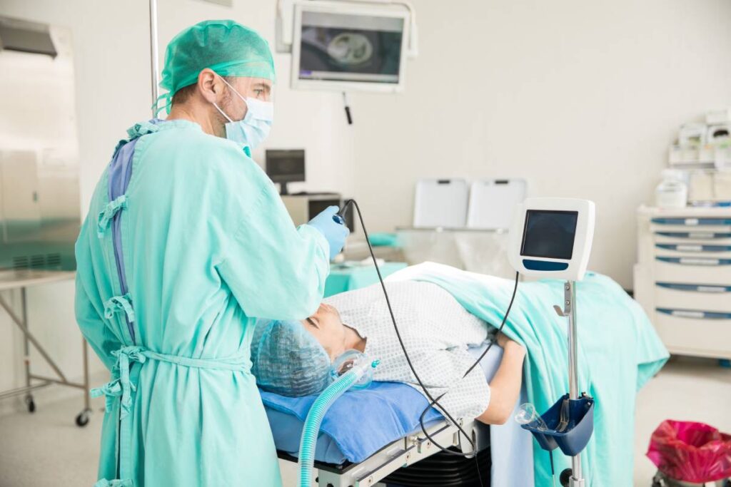
Video laryngoscopes, on the other hand, incorporate a camera positioned in the distal half of the blade that transmits images to a screen. This allows the clinician to view the vocal cords on the screen (indirect laryngoscopy) and guide the endotracheal tube through the vocal cords without a direct line of sight from the mouth. Video laryngoscopes offer advantages such as improved glottic visualization and potentially higher first-attempt success rates compared to traditional direct laryngoscopy. Video laryngoscopes come in various blade shapes and with or without tube advancement channels. Incorporating fiberoptic, video, optical, and mechanical technologies, these devices aid in managing difficult airways or facilitating intubation in adults, with many suitable for pediatric use as well.
The choice between direct laryngoscopy and video laryngoscopy depends on various factors, including the clinician’s experience and patient characteristics. Direct laryngoscopy is most easily performed with the clinician standing at the patient’s head and the bed adjusted to the level of the clinician’s xiphoid. Video laryngoscopes have become increasingly preferred based on data showing improved success metrics.
Observational studies comparing video laryngoscopy and direct laryngoscopy for pediatric intubation have found video laryngoscopy associated with similar or reduced adverse effects and similar or higher first attempt success rates. For instance, a cohort study of over 1400 pediatric emergency intubations showed that video laryngoscopy was associated with higher odds of first-attempt success and decreased odds of severe adverse airway outcomes compared to direct laryngoscopy. Similarly, in pediatric intensive care units, the implementation of routine video laryngoscopy with coaching led to a significant increase in video laryngoscopy usage during intubations and lower adverse event rates compared to direct laryngoscopy. Another multicenter study found similar first-pass success rates for video laryngoscopy and direct laryngoscopy in children, though video laryngoscopy was used for the entire procedure in only a minority of cases.
Confirming proper placement of the endotracheal tube is crucial and can be done using colorimetric end-tidal carbon dioxide devices or capnographic monitors. These devices provide accurate confirmation of endotracheal intubation and should be used in any setting where intubation is performed.
Endotracheal intubation is a complex procedure that requires careful planning, assessment, and the use of appropriate equipment. Video laryngoscopes offer advantages over traditional direct laryngoscopy, particularly in difficult airway cases, and can improve the success and safety of the procedure. Confirmation of proper tube placement is essential to ensure the patient’s airway is protected and oxygenated adequately.
References
Hansel J, Rogers AM, Lewis SR, Cook TM, Smith AF. Videolaryngoscopy versus direct laryngoscopy for adults undergoing tracheal intubation. Cochrane Database Syst Rev. 2022 Apr 4;4(4):CD011136. doi: 10.1002/14651858.CD011136.pub3. PMID: 35373840; PMCID: PMC8978307.
Ford HR, Gardner MJ, Lynch JM. Laryngotracheal disruption from blunt pediatric neck injuries: impact of early recognition and intervention on outcome. J Pediatr Surg. 1995 Feb;30(2):331-4; discussion 334-5. doi: 10.1016/0022-3468(95)90584-7. PMID: 7738760.
Kendall JL, Anglin D, Demetriades D. Penetrating neck trauma. Emerg Med Clin North Am. 1998 Feb;16(1):85-105. doi: 10.1016/s0733-8627(05)70350-3. PMID: 9496316.
Verghese ST, Hannallah RS. Pediatric otolaryngologic emergencies. Anesthesiol Clin North Am. 2001 Jun;19(2):237-56, vi. doi: 10.1016/s0889-8537(05)70227-3. PMID: 11469063.
Holm-Knudsen R. The difficult pediatric airway: a review of new devices for indirect laryngoscopy in children younger than two years of age. Paediatr Anaesth. 2011 Feb;21(2):98-103. doi: 10.1111/j.1460-9592.2010.03487.x. Epub 2010 Dec 15. PMID: 21159025.
Oakes ND, Dawar A, Murphy PC. Difficulties using the C-MAC paediatric videolaryngoscope. Anaesthesia. 2013 Jun;68(6):653-4. doi: 10.1111/anae.12298. PMID: 23662770.
Owada G, Mihara T, Inagawa G, Asakura A, Goto T, Ka K. A comparison of the Airtraq®, McGrath®, and Macintosh laryngoscopes for difficult paediatric intubation: A manikin study. PLoS One. 2017 Feb 10;12(2):e0171889. doi: 10.1371/journal.pone.0171889. PMID: 28187213; PMCID: PMC5302788.