Fiberoptic intubation is a specialized technique used to manage difficult airway situations where traditional intubation methods may be inadequate or impossible. This technique utilizes a flexible, thin tube fitted with a fiberoptic camera to provide a real-time, clear view of the airway, allowing for precise placement of an endotracheal tube. It has become an invaluable tool in cases involving anatomical abnormalities, trauma, or other complications that complicate airway management.
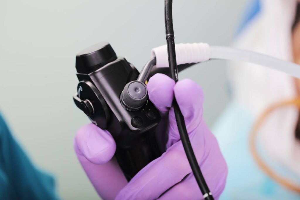
At the heart of fiberoptic intubation is the fiberoptic bronchoscope, a flexible instrument that incorporates a bundle of optical fibers. This design allows the clinician to see a magnified view of the airway structures, including the larynx, trachea, and bronchi, on a monitor. This visualization is critical for guiding the endotracheal tube through the vocal cords and into the trachea accurately.
During the procedure, the patient is typically sedated and prepared with local anesthesia to minimize discomfort. The bronchoscope is gently inserted through the nose or mouth, depending on the patient’s anatomy and clinical condition. As the bronchoscope advances, the clinician views the airway on a monitor, carefully maneuvering the scope into the correct place and positioning the endotracheal tube. Once the tube is correctly placed, it is secured, and the bronchoscope is removed.
Fiberoptic intubation is particularly useful in several scenarios. It is an excellent option for patients with difficult airway management needs, such as those with limited neck mobility, severe obesity, or facial deformities. Traditional intubation techniques may struggle in these cases, but fiberoptic intubation provides a direct view of the airway, allowing for more precise navigation. It is also beneficial in trauma situations, where facial or neck injuries can distort anatomy, making conventional intubation challenging. Fiberoptic intubation enables clinicians to bypass these obstructions by visualizing the airway structures directly and guiding a flexible device through the airway.
In cases where patients have known or suspected airway obstructions, such as tumors or severe inflammation, fiberoptic intubation allows for both assessment and management of these obstructions. Additionally, this technique can be performed with the patient awake if necessary, which is especially useful when full sedation is not advisable. Local anesthetics are used to numb the airway, allowing the patient to remain conscious while the procedure is carried out.
Despite its advantages, fiberoptic intubation does have limitations. It requires significant skill and experience, as mastery of the technique is not easily acquired. Inexperienced clinicians may face difficulties, particularly in high-pressure situations. Additionally, some patients with severe airway inflammation or other complicating factors may not be ideal candidates for this method.
In summary, fiberoptic intubation is an important component of airway management, offering a sophisticated and effective solution for challenging intubation scenarios. By providing a detailed view of the airway, this technique enhances the safety and efficacy of intubation procedures. Although it requires specialized training and resources, its ability to navigate complex anatomical challenges makes it an essential tool in modern medical practice. Continuous training and practice are vital to ensuring that clinicians can achieve the best possible outcomes for their patients.