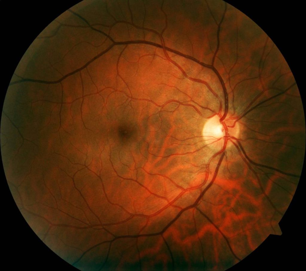
During rhegmatogenous retinal detachment (RRD) surgery, operating teams reattach a split sensory retina to the retinal pigment epithelium [1]. During the procedure, the anesthesiologist must ensure the safety of the patient and the comfort of the surgeon(s) [2]. From weighing regional versus general techniques to determining the best method through which to administer anesthesia, anesthesiologists have much to consider when choosing the optimal approach for retinal detachment surgery.
Before the 1990s, general anesthesia was the primary anesthetic used during RRD surgery [1]. Since then, regional anesthesia has become far more common [1]. This is because it avoids complications associated with general anesthesia (most notably, nausea and vomiting), while being cost-effective and promoting faster recovery [1].
However, these trends are not definitive: general anesthesia could be preferable in some cases [2]. For instance, obese patients at risk of airway loss may benefit from general anesthesia over regional anesthesia [2]. Additionally, regional anesthesia can result in inflammation, tissue division, repeated ocular muscular traction, and cryopexy [4]. General anesthesia could allow patients to avoid experiencing these forms of discomfort; alternatively, eye blocks could be administered alongside ketorolac, an analgesic comparable to morphine [4]. Therefore, anesthetists should assess patients’ preexisting conditions, airway complexity, and cardiopulmonary status to arrive at personalized anesthesia plans [2].
Peribulbar and retrobulbar blocks are the most common regional anesthetic options during RRD repair. While both means of regional anesthesia are considered relatively safe, peribulbar anesthesia is generally considered less risky than retrobulbar anesthesia [5]. This is because of the heightened chance of scleral perforation associated with the latter block [5]. When scleral perforation occurs, patients report experiencing a sharp pain, followed by a loss of vision, which often must be corrected by surgical intervention [5]. Other risks associated with retrobulbar injections include proliferative vitreoretinopathy, vitreous hemorrhage, retinal tear, retinal toxicity, and choroidal hemorrhage [5]. Anesthetists should take all of these risks into consideration when deciding between peribulbar and retrobulbar blocks.
Despite its relative risks, retrobulbar anesthesia has higher rates of efficacy than peribulbar anesthesia, so the choice between either technique is not always clear-cut. A case study analyzing an incidence of globe penetration during retrobulbar anesthesia suggested that, upon observing penetration, timelier detection and response help correct damage [5]. Of course, such adverse occurrences do not occur often. Given that globe penetration only occurs in about 0.075% of cases, retrobulbar block still merits consideration for RRD surgery patients [5].
Besides peribulbar and retrobulbar blocks, sub-Tenon anesthesia is another option that anesthesiologists can turn to during RRD repair. While the aforementioned blocks are most common, the sub-Tenon block is rising in popularity due to its reduced risks [6]. Because it is an episcleral technique, sub-Tenon avoids the complications associated with sharp needles passing through the ocular orbit [6]. Unfortunately, no light perception can occur at several points during surgery when patients are under sub-Tenon’s anesthesia, suggesting that the block may restrict optic nerve conduction [7]. Regardless, the harmlessness and transience of this condition suggest that sub-Tenon is still safe to use [7].
Ultimately, when choosing between different anesthetic methods for retinal detachment surgery, anesthetists should consider the complications associated with each type of anesthetic alongside a patient’s unique needs and comorbidities. The best choice will minimize the risk of events such as perforation and pain while enabling the most successful outcomes.
References
[7] W. Q. Chen et al., “Visual impact of sub-tenon’s anesthesia during surgery for retinal detachment,” Chinese Journal of Ophthalmology, vol. 53, no. 5, p. 332-337, May 2017. [Online]. Available: https://doi.org/10.3760/cma.j.issn.0412-4081.2017.05.004.
[5] Y. Dai, T. Sun, and J.-F. Gong, “Inadvertent globe penetration during retrobulbar anesthesia: A case report,” World Journal of Clinical Cases, vol. 9, no. 8, p. 2001-2007, March 2021. [Online]. Available: https://doi.org/10.12998/wjcc.v9.i8.2001.
[2] R. B. Singh et al., “Ocular complications of perioperative anesthesia: a review,” Graefe’s Archive for Clinical and Experimental Ophthalmology, vol. 2021, p. 1-15, February 2021. [Online]. Available: https://doi.org/10.1007/s00417-021-05119-x.
[4] X. Chen et al., “The Combination of Ketorolac with Local Anesthesia for Pain Control in Day Care Retinal Detachment Surgery: A Randomized Controlled Trial,” Journal of Ophthalmology, vol. 2017, p. 1-8, July 2017. [Online]. Available: https://doi.org/10.1155/2017/3464693.
[3] S. Riaz et al., “Shifting Paradigm: From General Anesthesia to Local Anesthesia in Posterior Segment Surgeries,” Journal of Ophthalmology, vol. 36, no. 3, p. 263-266, May 2020. [Online]. Available: https://doi.org/10.36351/pjo.v36i3.1001.
[1] R. Malagola et al., “Shifting Paradigm: From General Anesthesia to Local Anesthesia in Posterior Segment Surgeries,” Giornale di Chirugia, vol. 39, no. 4, p. 227-231, July-August 2018. [Online]. Available: https://bit.ly/3hh4CPN.
[6] P. Guise, “Sub-Tenon’s anesthesia: an update,” Local and Regional Anesthesia, vol. 2012, no. 5, p. 35-46, June 2012. [Online]. Available: https://doi.org/10.2147/LRA.S16314.
